-40%
pain management c-arm fluoroscopy xray siemens orbic
$ 8799.64
- Description
- Size Guide
Description
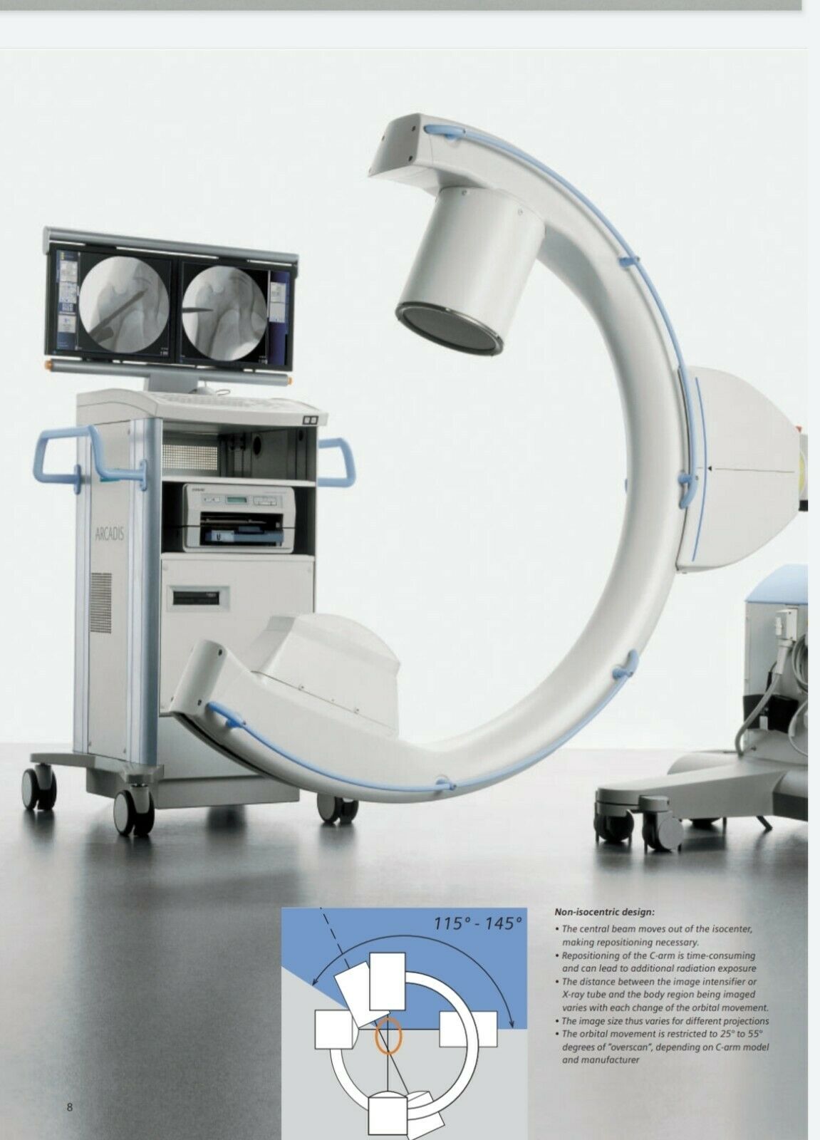
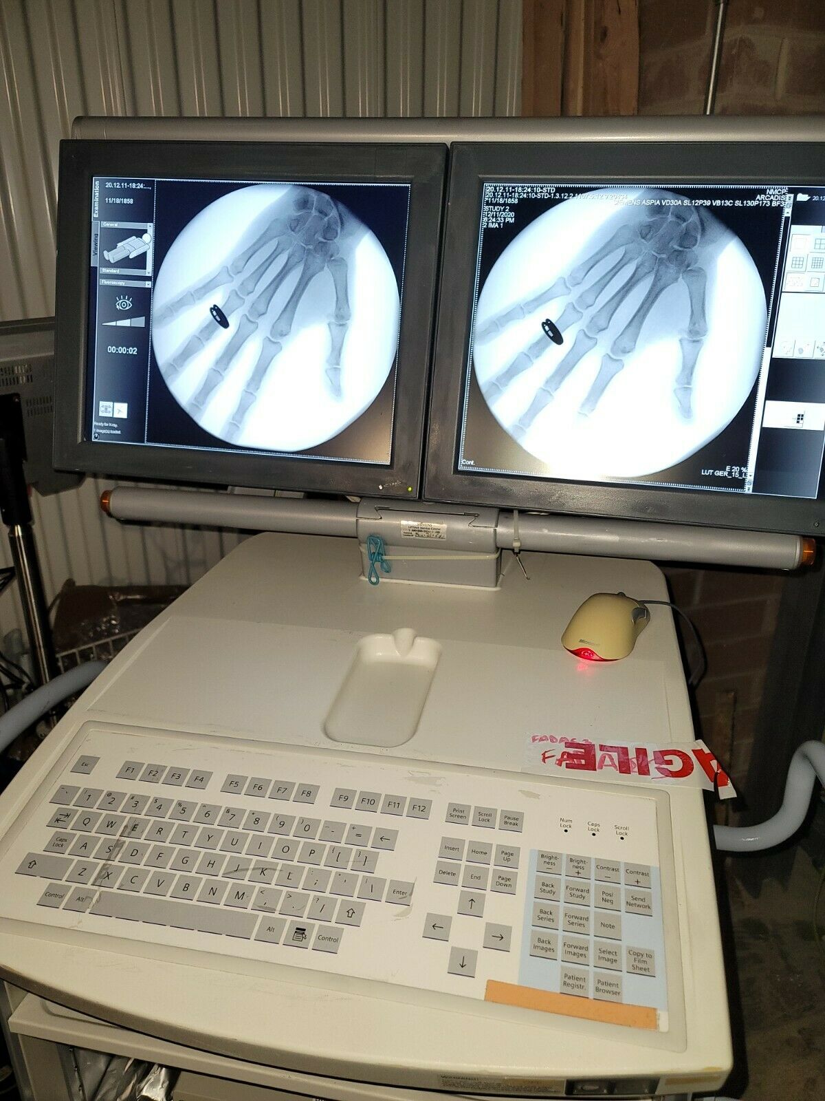
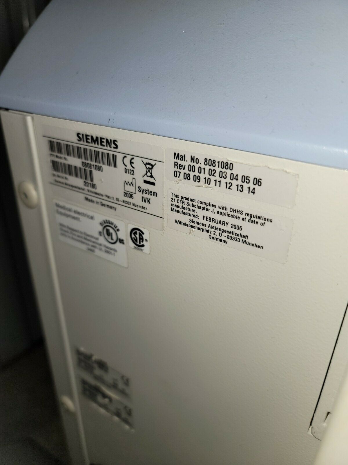
2006 C-Arm with 2015 newest model workstation computer M720Siemens Arcadis
Specifications
C-arm:
Isocentric C-arm movement with integrated cables and electromagnetic brakes
Orbital movement 190° (± 95°)
Angulation ± 190°
Horizontal movement 20 cm
Immersion depth 73 cm
Swivel range ± 10°
Vertical travel 40 cm
Source – I.I. distance 98 cm
Free space 78 cm
X-Ray Generator/Tube:
Max. pulsed output 2.3 kW
Inverted control frequency 15 kHz to 30 kHz
KV range 40 kV to 110 kV
Fluoroscopy 0.2 mA to 15.2 mA (max. 1000W)
Digital radiography 0.2 mA to 23 mA (max. 1000W)
Pulsed fluoroscopy Up to 23 mA
Pulse width Min. 7 ms
Pulse rate Up to 8 f/s, up to 15 f/s
Power mode enables temporary max. output in continuous fluoroscopy and pulsed fluoroscopy
Single Tank with Stationary Anode:
Focal spot nominal value 0.6
Nominal voltage 110 kV
Optical anode angle 9°
Inherent filtration ≥ 3 mm AI equivalent
Collimator System:
Iris diaphragm for concentric, radiation-free collimation
Semi-transparent slot diaphragm for symmetric, radiation-free collimation, with unlimited rotation
X-Ray TV System:
High-resolution X-Ray TV system in maintenance-free CCD technology with 1024 x 1024 (1k
2
) full-size CCD sensor
Constant image brightness due to automatic gain control
High contrast and high spatial resolution
TV matrix 1k
2
Digital image rotation ± 360°
X-Ray Image Intensifier:
Metal-enamel technology with Mu-metal shielding
Precision electron optics with minimal image distortion and consistent high resolution across the entire image field
Cesium-iodide input screen for minimum quantum noise and excellent resolution
Anti-glare output screen with scattered light trap for high contrast dynamics and prevention of scattered light effects
High-transparency input window
Nominal diameter 23 cm
Zoom format 15 cm
Grid PB 17/70, f
o
100
Trolley Options
19″ TFT Monochrome Display:
Screen size 48 cm (19″)
Image matrix 1280 x 1024
Brightness, typical 400 cd/m
2
Maximum brightness, typical 600 cd/m
2
Horizontal/vertical viewing angle 170°/170°
High-contrast display with high luminance
19″ TFT Color Display:
Screen size 48 cm (19″)
Image matrix 1280 x 1024
Brightness, typical 200 cd/m
2
Maximum brightness, typical 250 cd/m
2
Horizontal/vertical viewing angle 170°/170°
Column Display Flex:
Allows for vertical display positioning independently of trolley position with a rotation angle of -30° to 180°
Defined lock in place positions at 0° and 180°
Column Display Flex Plus:
Allows for vertical display positioning independently of trolley position with a rotation of 0° to 180°
Motorized height adjustment
Protection and transportation management by foldable displays
Defined lock in place positions at 0°, 90° and 180°
Sound System – Emotion:
Onboard sound system in order to connect audio devices via aux-in connector
Stereo amplifier with volume controller
Two blinded oval stereo loudspeakers
3D Image Acquisition:
Motor Drive
Motor-driven orbital movement through 190° for online reconstruction of isotropic 3D data sets
Rotation time
30 seconds with 50 images/scan
60 seconds with 100 images/scan
3D Visualization:
The following visualization modes are possible:
Multiplanar Reconstruction (MPR)
– With MPR, two-dimensional images of arbitrary orientation (axial, sagittal, coronal, oblique, double-oblique, curvilinear) are reconstructed from the isotropic 3D volume
Surface Shaded Display (SSD)
– This method is especially suitable for displaying bone surfaces. The reconstruction threshold value can be determined arbitrarily, resulting in surface reconstructions with special shading effects. The viewing angle and location can be freely selected
Volume Rendering Technique (VRT)
– Direct VRT allows highly precise, CT-like 3D visualization and easy orientation in the dataset
3D Image Fusion
– 3D Image Fusion for the spatial orientation and visualization of a patient’s image data that was generated by different modalities at different times. Supports optimal diagnosis (fusion of morphological) and functional information, therapy planning and follow-up
3D Dual-Monitor Display
– Is an extended feature for 3D and facilitates the viewing and manipulation of two different datasets on two monitors (monitor A and monitor B).
It allows the user to compare two studies from the same patient, i.e., pre-and intra-operative, or two studies from the same patient, i.e., pre and post-surgery with synchronized scrolling through both data sets
About xraychicago Management
Our
MARKET
is for used and new radiology equipment. We buy, sell, consign, and repair radiology equipment.
Our comprehensive services can help you clear space, reclaim capital, replace equipment, and repair almost anything radiology related. We can even help you remotely or come onsite.
please ask questions : [email protected]
Legal Information
Sale includes only items and accessories explicity shown or listed in the text of this advertisement. Additional accessories, features, and items are not included. we warrant the item to be in the condition described, but makes no guarantee regarding compatibility with other equipment, software, or use other than the item's original intended use.
This agreement will be governed by the laws and any disputes arising from this agreement shall be decided exclusively by the district or magistrate state courts in the Illinois. Any party that unsuccessfully challenges the enforceability of this Choice of Law and Forum Selection clause shall reimburse the prevailing party for its attorney fees and costs associated with defending against the challenge of said clause.
All devices are thoroughly cleaned to manufacturer specifications and inspected before listed for sale.
The sale of this item may be subject to strict regulation by the US Food and Drug Administration and state and local regulatory agencies. If so, do not bid on this item unless you are an authorized purchaser. If the item is subject to state or FDA regulation, you must provide verification that you are an authorized user.
